

Several aspects of Applied Physics are under development within the Department of Physics, in particular Environmental Acoustics and Medical Physics.
Environmental Acoustics
Environmental Acoustic research focuses on the generation and propagation in the environment of those sounds that can be dangerous for living beings. It studies methods for source identification and reduction of their emitted power, propagation models and insulation at the receiver. It makes use of forefront innovative instruments like an acoustic camera with 112 microphones or machine learning methods for source classification. These innovative tools allows researchers to deal with issues like port noise, not yet characterized.
Medical Physics
Research in Medical Physics deals with developing the instrumentation and analysis methods necessary to create cutting-edge systems. An imaging system is composed of a radiation-sensitive element followed by the relative readout and acquisition electronics (DAQ), and by an image processing and reconstruction software. For the development of prototypes that go beyond the state of the art, high-performance components must be used, integrated and optimized with each other. The use of innovative techniques for image analysis also contribute to the improvement of the performance of an imaging system, and enables advances in diagnostics and therapy.
All the skills needed for these developments are present in our Department:
- Nuclear imaging, and PET in particular: studies on scintillators, starting from the characterization of the spatial, temporal and energetic resolution properties of scintillating materials up to the evaluation of the different crystal structures (segmented, continuous and composite for the detection of the interaction depth) and on the characteristics of silicon photodetectors and optimization of their properties. A detecting module for ultra-time-of-flight PET detectors with on-chip neural network enhanced signal processing, featuring a CTR<200 ps, an intrinsic spatial resolution <1 mm, and 11% of energy resolution, has been developed in the H2020 UTOFPET project. The study with the innovative detecting module will continue, to enhance its characteristics and also thanks to its scalability, it is planned to develop systems that will be applied in small animal imaging, and total-body PET.
- In strict collaboration with final users, e.g., the Institute of Clinical Physiology of CNR (IFC), AOUP, Fondazione Toscana “Gabriele Monasterio” or international high-tech companies, we develop multi-modal imaging systems, such as preclinical PET/CT scanners. This is a highly interdisciplinary activity, including not only hardware but also software such as 3D image reconstruction algorithms (iterative such as ML-EM for PET and analytical such as filtered backprojection for CT) and acquisition and data calibration tools that are necessary for the final applications. The group also collaborates to the final clinical or preclinical applications during the preparation and execution of experiments. Part of these activities are performed in the framework of the Multi-Modal Molecular Imaging Italian node of Eurobioimaging, which is an ERIC (European Research Infrastructure Consortium) providing access to advanced services for biological and biomedical research. The Medical Physics group is also working on the implementation, in collaboration with private companies and the AOUP, of a trimodal system PET-MR-EEG (EU FP7 TRIMAGE project).
- Systems for 2D and 3D X-ray imaging, based on pixelated semiconductor detectors (Si and CdTe) bump-bonded to an ASIC, Timepix4, and readout by a dedicated acquisition system. This system was developed inside the international collaboration MEDIPIX, participated by INFN. This imaging system will be applied both in the pre-clinical and clinical fields in collaboration with other University departments, IFC and AOUP on applications such as dosimetry, 2D and 3D spectral imaging.
- Magnetic Resonance Imaging in collaboration with the IRCCS Stella Maris and its IMAGO7 Research Center, where there are clinical and ultra-high-field tomographs that operate on humans. Research includes: the development of different MR imaging methods through the investigation of different aspects such as detection techniques, pulse sequence design, data processing, imaging analysis, computational modeling, development of classification algorithms, and finally their application in vivo, either in field of neuroscience or in the understanding of physio-pathological mechanisms of different diseases.
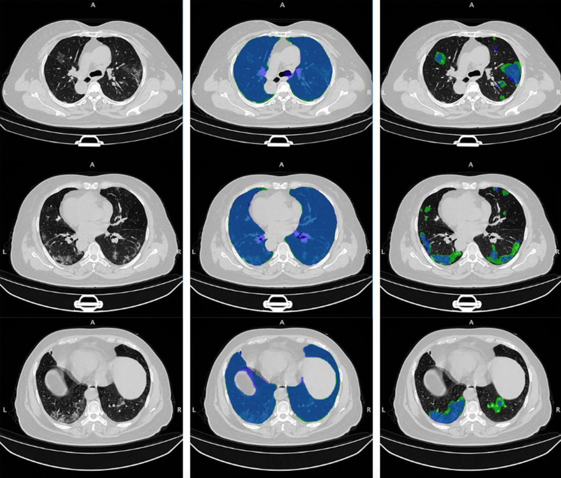
- Development and clinical validation of data analysis techniques based on Artificial Intelligence, including machine learning and deep-learning approaches, to foster their implementation in medical diagnostics and therapy. Current challenges include a variety of data mining, image segmentation, annotation and analysis applications, strategies for data harmonization, identification of quantitative image features, to realize intelligent systems for dosimetric applications, computed-assisted diagnosis and image-guided therapy.
- PET systems dedicated to the monitoring of treatment plans in radiotherapy with charged particles, collaborating with the INFN and with the main Italian and European centers for the experimentation of PET systems and for the development of new instrumentation beyond the state of the art. The developed systems are used for basic studies (DoPET) and for clinical studies: a clinical trial is underway at CNAO using INSIDE, a system that includes an in-beam PET system to observe the morphological changes undergone by the tumor mass during therapy and if necessary, update the treatment plan. INSIDE is a collaboration of the Department of Physics, INFN and CNAO.
- Nuclear reactions occurring in the patient during the delivery of a treatment plans in radiotherapy with charged particles, are the subject of research aiming to measure the cross sections of nuclear fragmentation reactions. This study takes place thanks to an international collaboration, FOOT, made up of 15 institutions and funded by INFN. FOOT is an experiment consisting of several sub-detectors, and the Medical Physics group coordinates the experimental activities dedicated to the identification of charged nuclear fragments. The experiment will take data at GSI in Germany and at CNAO in Pavia. For the hadrontherapy applications and related experiments the group is part of the Biophysics International Collaboration based at GSI.
- Development of new technologies for radiotherapy and in particular for FLASH radiotherapy, an advanced technique in which the radiation dose (few Gy’s) is delivered in less than a second unlike conventional radiotherapy in which the treatment is fractionated in several sessions of few minutes each. In the FLASH mode, with the same effect on the tumor, a significant decrease in the toxicity of the treatment was observed in the healthy tissues crossed by the radiation thus opening new scenarios for the cure of tumors hitherto untreatable. Numerous studies are underway to effectively and safely translate this radiobiological effect into clinical practice. For this reason, the Multidisciplinary Pisa Center on Research and Clinical Implementation of Flash Radiotherapy (CPFR) has been created, which involves the University of Pisa, the Pisa University Hospital AOUP, the CNR–Neuroscience Institute, and INFN. The medical physics group played a leading role in the foundation of the center and is currently involved in the development of new instrumentation and dosimetry protocols necessary for the accurate measurement of the radiation dose in ultra-intense regimes.
Last updated on: 30/03/2023